Stent-Assisted Detachable Coil Embolization of Two Splenic Artery Aneurysms
Paulo Eduardo Ocke Reis and Guilherme de Palma Abrão
DOI10.36648/2634-7156.21.6.11
Paulo Eduardo Ocke Reis1,2* and Guilherme de Palma Abrão3
1Department of Specialized and General Surgery, Universidade Federal Fluminense, Rio de Janeiro, Brazil
2Vascular Clinic Ocke Reis, Rio de Janeiro, Brazil
3Department of Radiology, Universidade Federal Fluminense, Rio de Janeiro, Brazil
- *Corresponding Author:
- Paulo Eduardo Ocke Reis
Department of Specialized and General Surgery, Fluminense Federal University, Rio de Janeiro, Brazil
Tel: +55 21 2629-5000
E-mail: vascular@pauloocke.com.br
Received Date: January 28, 2021; Accepted Date: March 23, 2021; Published Date: March 30, 2021
Citation: Ocke Reis PE, Guilherme de PA (2021) Stent-Assisted Detachable Coil Embolization of Two Splenic Artery Aneurysms. J Vasc Endovasc Ther. 6 No. 3: 11.
Image Article
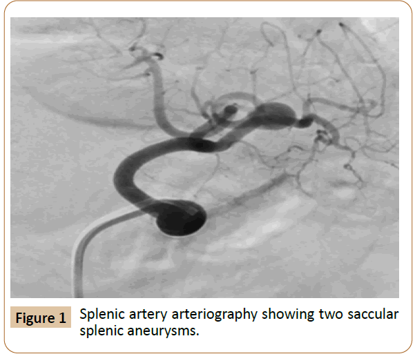
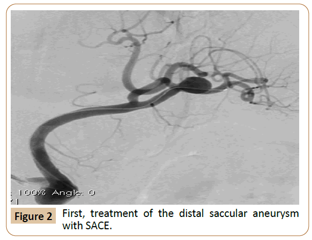
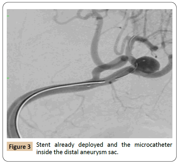
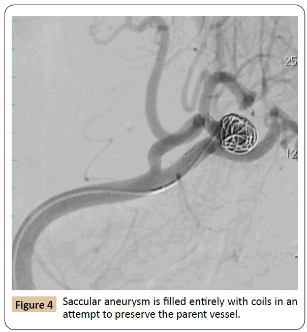
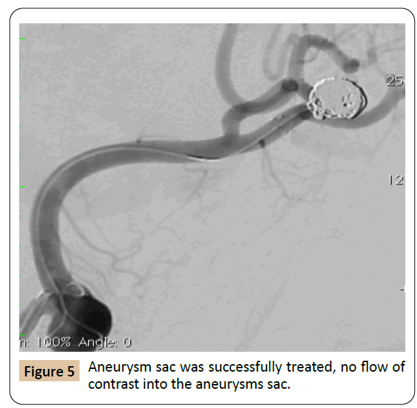
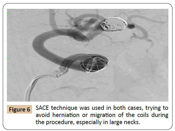
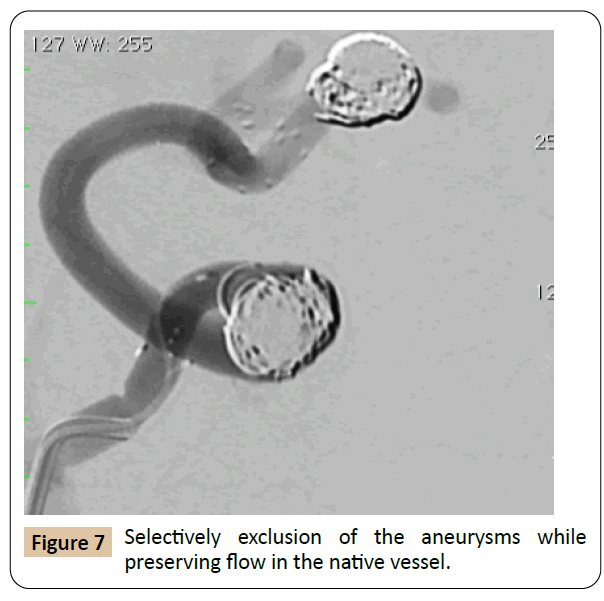
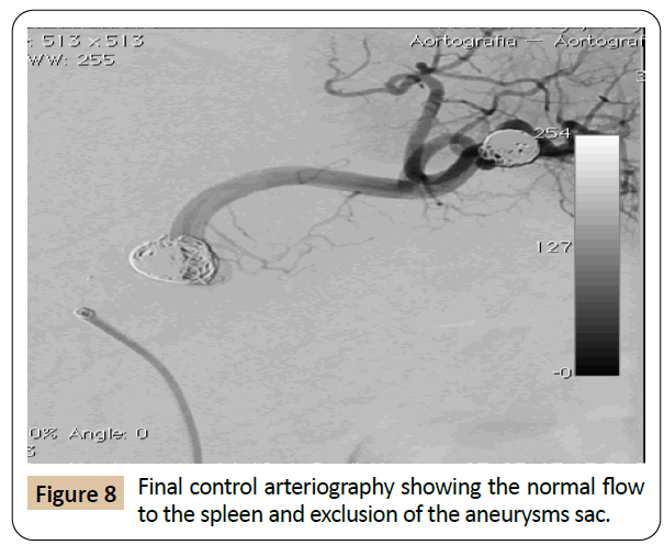
Visceral artery aneurysms (VAAs) challenging cases, as widenecked aneurysms using stent assisted coil embolization (SACE),to treat two splenic aneurysms. The goal of treatment is to prevent aneurysm expansion by excluding it from the arterial circulation, saving branches, patency, and freedom from rupture or reperfusion [1-6].
Step by Step
References
- Cordova AC, Sumpio BE (2013) Visceral artery aneurysms and pseudo aneurysms-should they all be managed by endovascular techniques? Ann Vasc Dis 6: 687-693.
- Hemp JH , Sabri SS (2015)Endovascular management of visceral arterial aneurysms. Techniques Vascular and Interventional Radiology 18: 14-23.
- Ocke Reis PE, Roever L, Ocke Reis IF, De Azambuja Fontes F, Rotolo Nascimento M, et al. (2016) Endovascular stent grafting of a deep femoral artery pseudo aneurysm. EJVES Short Rep 26:5-8.
- Mkangala AM, Liang H, Dong XJ, Su Y, HaoHao L (2020) Safety and efficacy of conservative, endovascular bare stent and endovascular coil assisting bare stent treatments for patients diagnosed with spontaneous isolated superior mesenteric artery dissection. WideochirInne Tech Maloinwazyjne 15:608-619.
- Bi G, Xiong G, Dai X, Shen X, Deng L (2020) Endovascular repair of an aneurysm arising from a celiomesenteric trunk. Ann Vasc Surg 62:497.
- Ocke Reis PE, Roever L, Reis IFO (2016) Embolization for visceral artery aneurisms: what’s your opinion? J Vasc Endovasc Surg 1:1.
Open Access Journals
- Aquaculture & Veterinary Science
- Chemistry & Chemical Sciences
- Clinical Sciences
- Engineering
- General Science
- Genetics & Molecular Biology
- Health Care & Nursing
- Immunology & Microbiology
- Materials Science
- Mathematics & Physics
- Medical Sciences
- Neurology & Psychiatry
- Oncology & Cancer Science
- Pharmaceutical Sciences
