A Mycotic Radial Artery Pseudoaneurysm in the Anatomical Snuff Box: A Case Report
Mokhtari S, Abutabeekh N, Abualtayef T, Oulad amar A, Kamaoui I,Benzirar A and Elmahi O
DOI10.21767/2573-4482.19.04.21
Mokhtari S1*, Abutabeekh N1, Abualtayef T1, Oulad amar A2, Kamaoui I2, Benzirar A1 and Elmahi O1
1Vascular Surgery Department, Mohammed VI UHC, Oujda, Morocco
2Radiology Department, Mohammed VI UHC, Oujda, Morocco
- *Corresponding Author:
- Mokhtari S
Mokhtari S, Vascular surgery Department
Mohammed VI UHC, Oujda, Morocco
Tel: 639666566
E-mail:saramokhtari1904@gmail.com
Received date: September 09, 2019 Accepted date: September 20, 2019 Published date: September 27, 2019
Citation: Mokhtari S, Abutabeekh N, Abualtayef T, Oulad amar A, Kamaoui I, et al. (2019) A Mycotic Radial Artery Pseudoaneurysm in the Anatomical Snuff Box: A Case Report. J Vasc Endovasc Therapy 3:21
Abstract
Distal radial artery aneurysm of the hand is considered as a rare vascular pathology. Through this paper we report a case of a symptomatic snuff box radial artery pseudoaneurysm treated surgically by arterial ligation and aneurysm excision. The decision was based on a negative Allen’s test, an angio-CT and specially the constatation of an adequate back bleeding from the distal stump. Indeed, an accurate diagnosis and a preoperative blood flow evaluation are necessary for an appropriate management of distal radial artery aneurysms.
Keywords
Case report; Hemodialysis; Mycotic pseudoaneurysm; Anatomical snuff box; Vascular surgery; Radial artery
Introduction
Upper limb aneurysms are rare and represent only 1% of all locations. The involvement of the radial or ulnar artery is even rarer [1]. Two types of lesions are defined; true and false aneurysms.
Mycotic pseudoaneurysms of distal radial artery are uncommon disease. The majority of cases of the aneurysms in the snuff box been reported were due to traumatic, atheromatous causes or idiopathic.
This paper describes a rare case of 80 years old woman diagnosed with a mycotic pseudoaneurysm of radial artery in the anatomical snuff box. Through this case report and a recent extensive review of literature, clinical presentations, imaging tools, etiologies and therapeutic modalities being used in this pathology are determined and discussed.
Case Report
A 80-year-old woman, a chronic hemodialysis (3 sessions per week) was presented with a chief complaint of a swelling on the back of her right hand. A puncture and an aspiration of the mass content were performed at a private clinic. Due to the suspicion of an aneurysm, the patient was referred to our department for management. The history of the appearance of the mass was fuzzy. According to the patient, the mass has been discovered accidently just after a slight traumatism of the wrest; 4 days before being admitted in our department. No family history of aneurysms was identified in the anamnesis (medical record).
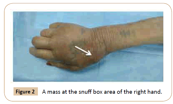
The physical examination revealed a painful pulsatile mass in the right anatomical snuff box with fragile skin and the puncture point scar facing. The peripheral pulses of upper members were perceived. Allen’s test was negative. There were no signs of other localization of arterial aneurysms. Laboratory examinations showed evidence of systemic inflammation (Leucocytosis: 21800; PCR: 300) without metabolic disorders or autoimmune diseases.
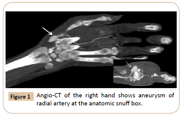
A Doppler ultrasound completed by an angio-CT showed a saccular pseudoaneurysm of palmar-radial artery that measures approximately 20mm. Considering the likelihood of aneurysm complications, especially the rupture in this case, a surgical intervention was performed urgently. Under loco-regional anesthesia, the aneurysm site was incised. An excision of the aneurysm and proximal and distal ligation of the artery were performed. The pathological and bacteriological examinations revealed a false aneurysm of mycotic cause. The post-operative follow-up was uneventful. The patient was discharged two days after vascular surgery under antibiotic treatment. The patient was symptom-free at two months follow-up (Figures 1-3).
Discussion
Upper extremity arterial aneurysms are rare, and radial artery aneurysms are even rarer [1,2]. The most common location of aneurysms of the hand is the distal ulnar artery [1]. Indeed, distal ulnar artery aneurysms have been well described as a clinical finding of hypothenar hammer syndrome [3] contrary to distal radial artery aneurysms of the hand, which are rarely reported and only in the form of case report. Until this day, we found 23 cases of radial artery aneurysms located in the anatomical snuff box in the English literature (Table 1) [4-24].
Table 1: Reported cases of radial artery aneurysm in the anatomical snuff box.
| Author (Year) | Age/Sex | Cause | Type of aneurysm | Treatment |
|---|---|---|---|---|
| Thorrens et al. (1966) [4] | 60/M | Arteriosclerosis | N.D | Excision and revascularization |
| Poirier and Stansel (1972) [5] | 69/M | Mycotic | N.D | Excision |
| Kleinert et al. (1973) [6] | 47/M | Trauma | False | Excision and revascularization |
| Kleinert et al. (1973) [6] | 53/F | Idiopathic | False | Excision |
| Malt (1978) [7] | 56/M | Arteriosclerosis | True | Excision and revascularization |
| Giler et al. (1979) [8] | 51/M | Buerger | N.D | Observation |
| Wenger et al. (1980) [9] | 60/M | Trauma | False | Excision |
| Wenger et al. (1980) [9] | 23/M | Trauma | False | Excision |
| Leitner et al. (1985) [10] | 69/F | Granulomatous arteritis | True | Excision |
| Walton and Choudhary (2002) [11] | 40/M | Idiopathic | N.D | Observation |
| Luzzani et al. (2006) [12] | 63/F | Idiopathic | True | Excision |
| Yaghoubian and de Virgilio (2006) [13] | 77/M | Idiopathic | N.D | Observation |
| Behar et al. (2007) [14] | 62/M | Trauma | True | Excision |
| Hegde et al. (2007) [15] | 55/F | Idiopathic | True | Excision |
| Umeda et al. (2009) [16] | 74/F | Marfan | True | Excision |
| Jadynak and Frydman (2012) [17] | 60/M | Idiopathic | True | Excision |
| H.I Shaabi (2014) [18] | 65/F | Idiopathic | True | Excision |
| Morin et al. (2014) [19] | 43/M | Trauma | False | Excision and revascularization |
| J.D Ayers (2015) [20] | 65/M | N.D | True | Excision and revascularization |
| Yamamoto et al. (2016) [21] | 61/F | Trauma | False | Excision |
| Nassiri et al. (2016) [22] | 16/M | N.D | False | Excision and revascularization |
| S.B Erdogan (2018) [23] | 52/M | Idiopathic | True | Excision and revascularization |
| Ghaffarian et al. (2018) [24] | 25/M | Idiopathic | True | Excision and revascularization |
| Our case (2019) | 80/F | Trauma | False | Excision |
N.D: Not described
The diagnosis of an aneurysm in the upper extremity can be easy in front of its typical clinical appearance by detection of a pulsatile mass. Some patients present with nonspecific symptoms ranging from an asymptomatic mass or a pain in the hand to distal ischemia, paresthesia, skin ulceration, and bleeding complications [25].
As the anatomical snuff box can be the seat of different pathologies like a ganglion, an abscess or a tumor, imaging modalities particularly duplex ultrasonography, CT angiography and MRI remain essential to confirm the diagnosis of an aneurysm and to guide the therapeutic management. Duplex ultrasonography can play a crucial role in distinguishing true arterial aneurysms from pseudoaneurysms by detecting the so-called “ying-yang” sign which is a pathognomonic of pseudoaneurysms cases [26]. In rare situations, pre-operative angiography can also be a valuable tool to help identify associated vascular pathologies (arteriovenous fistulas and malformations, fibromuscular dysplasia) and to identify the presence of collaterals.
Precise therapeutic options have been described for the management of this rare disease. It include careful observation, in case of small asymptomatic aneurysms [8,11,13]. Aneurysm excision with arterial ligation or arterial reconstruction, whether by end-end anastomosis or by vein graft in the case of aneurysmal dominant artery in order to supply the insufficient collateral blood being evaluated with a the preoperative blood flow measures. In our case, because of the high risk of rupture, the patient was urgently taken to the operating room where a surgical excision of the pseudoaneurysm and a ligation of radial artery were performed. The therapeutic decision was concluded due to the presence of adequate collateral flow and infection. A probabilistic antibiotic treatment was instituted secondarily adapted to the bacteriological result: a false aneurysm of mycotic origin. We performed a transthoracic ultrasound to look for endocarditis which could explain the distal artery mycotic pseudoaneurysm; however, it returned normal.
From a less invasive treatment point of view, endovascular therapeutic intervention using covered stent, coil and agent embolization are extremely limited alternative treatment option for radial artery aneurysms. It remains a controversial option [27].
Rupture of radial artery aneurysms has been rarely reported [28]. In our case, the rupture was favored by the fragility of the aneurysmal wall by infection and iatrogenic puncture. However, radial artery aneurysms are associated with the risk of thromboembolic events, distal ischemia, and nerve compression symptoms [21].
Published cases of radial artery aneurysm in the anatomical snuff box are attributed to trauma [6,9,14,19,21], atherosclerosis [4,7], mycotic [5], Marfan syndrome [16], systemic vasculitis [8,10] and idiopathic origin [6,11-13,15,17,18,23,24].
Conclusion
Radial artery aneurysms in the anatomical snuff box are exceedingly rare. They can lead to serious complications, reason why; in the majority of cases, their management remains necessary. Surgical excision of the aneurysm is the gold standard treatment option, the decision of performing revascularization requires an evaluation of the distal blood flow.
Conflict of Interest
No conflict of interest was declared by the authors.
References
- HO PK, Weiland AJ, McClinton MA, Wilgis EFS (1987) Aneurysms of the upper extremity. J Hand Surg 12: 39-46.
- Ogeng’o JA, Otieno B (2011) Aneurysms in the arteries of the upper extremity in aKenyan population. Cardiovasc Pathol 20: e53-e56.
- McClinton MA (2011) Reconstruction for ulnar artery aneurysm at the wrist. J Hand Surg 36: 328-332.
- Thorrens S, Trippel OH, Bergan JJ (1966) Arteriosclerotic aneurysms of the hand: excision and restoration of continuity. Arch Surg 92: 937-939.
- RA Poirier, HC Stansel Jr. (1972) Arterial aneurysms of the hand. Am J Surg 124: 72-74.
- Kleinert HE, Burget GC, Morgan JA, Kutz JE, Atasoy E (1973) Aneurysms of thehand. Arch Surg 106: 554-557.
- Malt S (1978) An arteriosclerotic aneurysm of the hand. Arch Surg 113: 762-763.
- Giler S, Zelikovski A, Goren G, Urca I (1979) Aneurysm of the radial artery in apatient with Buerger’s disease. VASA 8:147-149.
- Wenger DR, Boyer DW, Sandzén SC (1980) Traumatic aneurysm of the radialartery in the anatomical snuff box a report of two cases. Hand 12: 266-270.
- Leitner DW, Ross JS, Neary JR (1985) Granulomatous radial arteritis with bilateral,nontraumatic, true arterial aneurysms within the anatomic snuffbox. J Hand Surg 10: 131-135.
- Walton NP, Choudhary F (2002) Idiopathic radial artery aneurysm in theanatomical snuff box. Acta Orthop Belg 68: 292-294.
- Luzzani L, Bellosta R, Carugati C, Talarico M, Sarcina A (2006) Aneurysm of theradial artery in the anatomical snuff box. EJVES Extra 11: 94-96.
- Yaghoubian A, de Virgilio C (2006) Noniatrogenic aneurysm of the distal radialartery: a case report. Ann Vasc Surg 20: 784-786.
- Behar JM, Winston JS, Knowles J, Myint F (2007) Radial artery aneurysm resultingfrom repetitive occupational injury: tailor’s thumb. Eur J Vasc Endovasc Surg 34: 299-301.
- Ayers JD, Halbach J, Brown D (2015) True radial artery aneurysm: diagnosis and treatment. J Vasc Surg 62: 813-814.
- Umeda Y, Matsuno Y, Imaizumi M, Mori Y, Iwata H, et al. (2009) Bilateral radial artery aneurysms in the anatomical snuff box seen in Marfan syndrome patient: case report and literature review. Ann vasc dis 2: 185-189.
- Jedynak J, Frydman G (2012) Idiopathic true aneurysm of the radial artery: a rareentity. EJVES Extra 24: e21-e22.
- Shaabi HI (2014) True idiopathic saccular aneurysm of the radial artery. J Surg Case Rep 14: 1-3.
- Morina A, Della Schiavab N, Peyrachonc B, Collet-Benzaquend D, Longa A (2015) Post-traumatic aneurysm of the distal radial artery leading to digital ischemia. J des Mal Vasc 40 : 49-52.
- Hegde R, Addala P, Kumar S (2007) True Radial Artery Aneurysm in the Anatomical Snuffbox. Int J Surg 17: 15-17.
- Yamamoto Y, Kudo T, Igari K, Toyofuku T, Inoue Y (2016) Radial artery aneurysm in the anatomical snuff box: a case report and literature review. Int J Surg Case Rep 27: 44-47.
- Nassiri N, Kogan S, Truong H, Nagarsheth KJ, Shafritz R et al. (2016) Surgical repair of a snuffbox radial artery pseudoaneurysm. Clin Surg Vasc Surg 1: 1154.
- Erdogan SB, Akansel S, Selcuk NT, Aka SA (2018) Reconstructive surgery of true aneurysm of the radial artery: A case report. North Clin Istanb 5: 72-74.
- Ghaffarian AA, Brooke BS, Rawles J, Sarfati M (2018) Repair of a symptomatic true radial artery aneurysm at the anatomic snuff box with interposition great saphenous vein graft. J Vasc Surg Cases Innov Tech 4: 292-295.
- Dryton G, Allen KB, Borkon AM, Aggarwal S, Davis JR (2015) Do not bite the hand that feeds you. Ann Vasc Surg 29: 362-e3.
- Rozen G, Samuels DR, Blank A (2001) The to and fro sign: the hall-mark of pseudoaneurysm. Isr Med Assoc J 3: 781-782.
- Carrafiello G, Laganà D, Mangini M, Fontana F, Recaldini C, et al. (2011) Percutaneous treatment of traumatic upper-extremity arterial injuries: a single-center experience. J Vasc Interv Radiol 22: 34-39.
- Dawson J, Fitridge R (2013) Update on aneurysm disease: current insights and controversies: peripheral aneurysms: when to intervenedis rupture really a danger? Prog Cardiovasc Dis 56: 26-35.
Open Access Journals
- Aquaculture & Veterinary Science
- Chemistry & Chemical Sciences
- Clinical Sciences
- Engineering
- General Science
- Genetics & Molecular Biology
- Health Care & Nursing
- Immunology & Microbiology
- Materials Science
- Mathematics & Physics
- Medical Sciences
- Neurology & Psychiatry
- Oncology & Cancer Science
- Pharmaceutical Sciences


