Novel Technique to Cross Infrapopliteal Artery Chronic Total Occlusions
Bhaskar Purushottam and Prakash Krishnan
DOI10.21767/2573-4482.20.05.22
1Monument Health, Heart and Vascular Institute, Monument Health Rapid City Hospital, USA
2Mount Sinai Heart, Mount Sinai Medical Center, New York, USA
- *Corresponding Author:
- Bhaskar Purushottam
Monument Health, Heart and Vascular Institute
Monument Health Rapid City Hospital, USA
Tel: +16056465715
E-mail: bpurushottam@gmail.com
Received Date: August 14, 2020; Accepted Date: August 21, 2020; Published Date: August 28, 2020
Citation: Purushottam B, Krishnan P (2020) Novel Technique to Cross Infrapopliteal Artery Chronic Total Occlusions. J Vasc Endovasc Ther. 5 No. 4: 22
Abstract
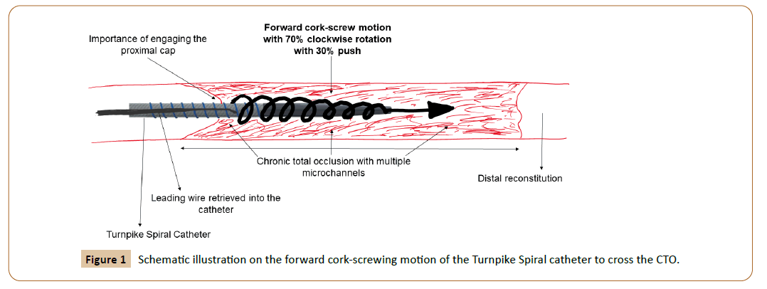
The standard endovascular approach to infrapopliteal artery chronic total occlusions [CTO] is the wire and catheter technique. In our paper we would like to describe a novel technique to cross these infrapopliteal artery CTOs using the Turnpike Spiral catheter. This technique involves a clockwise rotatory movement on the catheter with a very gentle forward push [70% rotation and 30% forward push]. In other words, the catheter is advanced in a forward screwing motion without a leading wire. We believe that the micro channels in these CTOs are the basis for our novel crossing technique.
Keywords
Critical limb ischemia; Support catheter; Spiraling; Cork-screw
Introduction
Patients with critical limb ischemia [CLI] commonly present with occlusive infrapopliteal arterial disease [1]. In most patients, endovascular route of treatment strategy is adopted for revascularization. A significant proportion of infrapopliteal arterial disease seen in CLI have chronic total occlusions [CTOs]. The standard approach to crossing these CTOs are wire escalation with a support catheter [2]. In addition to this standard approach there is a slew of specialty catheters and crossing devices [2,3] which add time and cost to the procedure.
Case Report
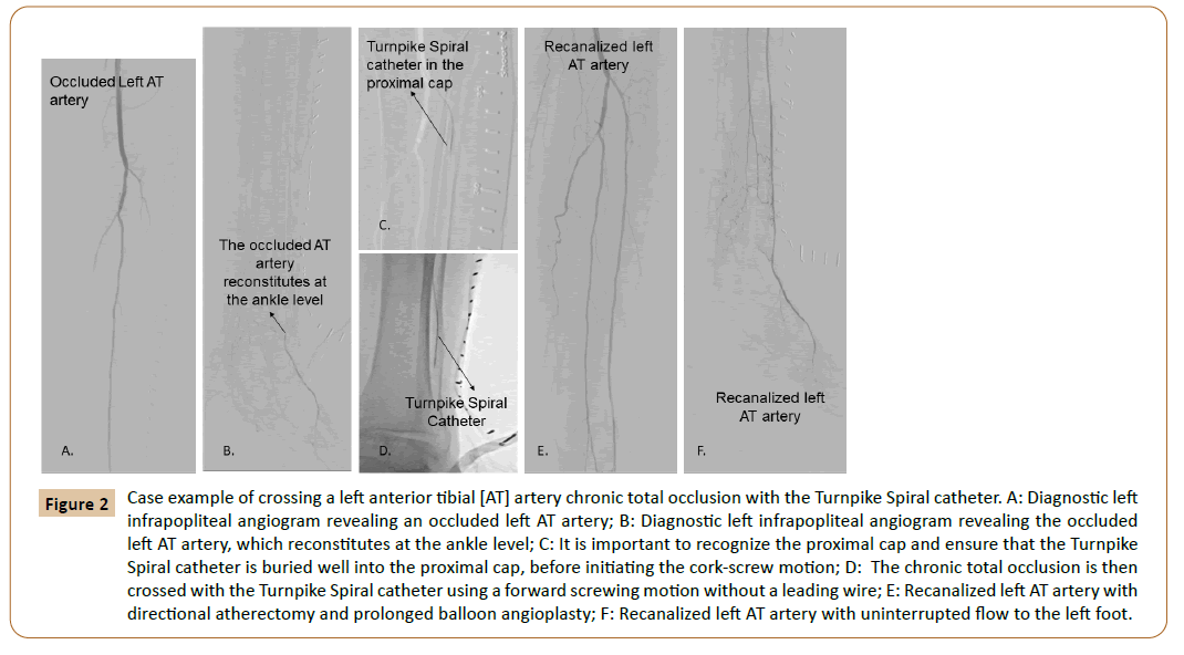
In this paper we would like to describe a new technique to cross infrapopliteal arterial CTOs using the Turnpike Spiral Catheter [Teleflex, USA] [4]. This catheter is approved as a support catheter for coronary CTOs. The company’s instructions for use manual recommends a leading wire over which the catheter is advanced. In this case report we describe a technique of advancing this catheter without a leading wire. After obtaining the diagnostic lower extremity angiograms and parking the guiding sheath in the mid femoropopliteal arterial segment, we obtain orthogonal views of the proximal cap of the infrapopliteal CTO. We would like to emphasize the fact it is very important to define the proximal cap of the CTO. Under roadmap fluoroscopic guidance, we introduce our work horse wire into the proximal cap of the CTO. We then advance the Turnpike Spiral catheter over the work horse wire and bury it into the proximal cap of the CTO. The wire is then retrieved into the catheter. We then perform a constant clockwise rotatory movement on the catheter with a very gentle forward push [70% rotation and 30% forward push]. In other words, the catheter is advanced in a forward screwing motion (Figure 1) without a leading wire. As we perform this movement, we assess the motion of the catheter tip, the tactile feedback and the direction it moves. If successful, the catheter transits through the body of the CTO with a distinctive tactile feedback. It is important to unwind the catheter in the opposite rotatory direction to avoid a buildup of torque (after performing clockwise forward rotatory movement of 10 turns, we then perform counterclockwise rotatory movement of 10 turns without pulling back the catheter). This way we gain adequate ground and prevent buildup of torque. When we approach the distal cap of the CTO, we obtain angiograms in orthogonal views. If satisfied with the direction of the catheter we then continue with the cork-screw motion and penetrate through the distal cap of the CTO. We then use the work horse wire to enter the distal lumen. If the wire does not sail through easily, we then obtain angiograms in orthogonal views to understand the distal reconstitution zone of the occluded artery and its relation to the catheter. At this point, we decide as to whether to proceed with the above described technique or switch to wire escalation/specialty crossing catheter/retrograde approach. If the direction of the catheter or tactile feedback feels extraluminal or in the presence of calcification, we abort the technique and proceed with other alternatives as described above. In some instances, if the catheter appears to be entering a potential branch or collateral, we retrieve the catheter back into the main arterial segment and then with the help of CTO wire, we guide the catheter into the appropriate course. A case example is depicted in Figures 2A-2F.
Figure 2: Case example of crossing a left anterior tibial [AT] artery chronic total occlusion with the Turnpike Spiral catheter. A: Diagnostic left infrapopliteal angiogram revealing an occluded left AT artery; B: Diagnostic left infrapopliteal angiogram revealing the occluded left AT artery, which reconstitutes at the ankle level; C: It is important to recognize the proximal cap and ensure that the Turnpike Spiral catheter is buried well into the proximal cap, before initiating the cork-screw motion; D: The chronic total occlusion is then crossed with the Turnpike Spiral catheter using a forward screwing motion without a leading wire; E: Recanalized left AT artery with directional atherectomy and prolonged balloon angioplasty; F: Recanalized left AT artery with uninterrupted flow to the left foot.
Discussion
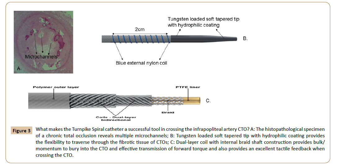
All chronic total occlusions have microchannels (Figure 3A) and this forms the basis for crossing these chronic total occlusions using the wire/support catheter technique and some crossing devices. We believe that these same microchannels favor our novel crossing technique. Given the inherent qualities of the Turnpike Spiral catheter as described below, the catheter tends to stay intraluminal once it enters the proximal cap of the CTO.
Figure 3: What makes the Turnpike Spiral catheter a successful tool in crossing the infrapopliteal artery CTO? A: The histopathological specimen of a chronic total occlusion reveals multiple microchannels; B: Tungsten loaded soft tapered tip with hydrophilic coating provides the flexibility to traverse through the fibrotic tissue of CTOs; C: Dual-layer coil with internal braid shaft construction provides bulk/momentum to bury into the CTO and effective transmission of forward torque and also provides an excellent tactile feedback when crossing the CTO.
The Turnpike Spiral catheter has the following key features, which makes it successful in crossing the CTOs:
• The proximal end has a sturdy base to help rotate the catheter (aids in the forward cork-screw motion).
• The crossing profile (distal tip outer diameter is 0.53 mm and the shaft out diameter is 0.97 mm) is ideal as infrapopliteal arteries have a diameter ranging from 1.5 mm to 4mm. If smaller, the catheter would not have the bulk to traverse the CTO. If larger, then it would lose the penetrating power.
This technique is successful when there is a well-defined cone shaped proximal cap, minimal calcification, absence of large collaterals and tortuosity. The main reason for failure is calcification. In the future, we intend to study this technique under transcutaneous ultrasound.
Conclusion
In summary, the Turnpike spiral catheter can be used as an alternative to cross infrapopliteal CTOs in the absence of significant calcification or tortuosity using a forward cork screwing motion, without a leading wire. The build of the catheter favors its course through microchannels and hence when successful, it almost always takes the intraluminal path. Therefore, we believe that this novel technique is an attractive alternative to crossing infrapopliteal artery CTOs as well as being cost-effective.
References
- Narula N, Dannenberg AJ, Olin JW, Bhatt DL, Johnson KW, et al. (2018) pathology of peripheral artery disease in patients with critical limb ischemia. J Am Coll Cardiol 72: 2152-2163.
- Kawarada O, Sakamoto S, Harada K, Ishihara M, Yasuda S, et al. (2014) Contemporary crossing techniques for infrapopliteal chronic total occlusions. J Endovasc Ther 21: 266-280.
- Singh GD, Armstrong EJ, Yeo KK, Singh S, Westin GG, et al. (2014) Endovascular Recanalization of infrapopliteal occlusions in patients with critical limb ischemia. J Vasc Surg 59: 1300-1307.
- https://teleflex.com/usa/en/product-areas/interventional/peripheral-interventions/turnpike-catheter/index.html
Open Access Journals
- Aquaculture & Veterinary Science
- Chemistry & Chemical Sciences
- Clinical Sciences
- Engineering
- General Science
- Genetics & Molecular Biology
- Health Care & Nursing
- Immunology & Microbiology
- Materials Science
- Mathematics & Physics
- Medical Sciences
- Neurology & Psychiatry
- Oncology & Cancer Science
- Pharmaceutical Sciences


