Wire Get Out of Right Foot after Enteral Venous Atheterisation
Reza Shojaee, Rohollah Davoodabadi, Melika Moazzami, Mohammadreza Rezaei and Mohamadreza Rohani
DOI10.36648/2634-7156.21.6.17
Reza Shojaee1, Rohollah Davoodabadi2, Melika Moazzami3, Mohammadreza Rezaei3 and Mohamadreza Rohani4*
1Vascular surgeon, department of surgery, assistant professor, Arak medical university, Iran
2Interventional cardiologist, assistant professor, Arak medical university, Iran
3Department of vascular surgery, researcher, Arak medical university, Iran
4Gasteroenterologist, assistant professor, Arak medical university, Iran
- *Corresponding Author:
- Mohamadreza Rohani
Gasteroenterologist, assistant professor, Arak medical university, Iran
Tel: 989123700960
E-mail: Mohamadreza.rohani@yahoo.com
Received Date: March 18, 2021; Accepted Date: April 06, 2021; Published Date: April 13, 2021
Citation: Reza S, Rohollah D, Melika M, Mohammadreza R, Mohamadreza R (2021) Wire Get Out of Right Foot after Enteral Venous Atheterisation. J Vasc Endovasc Therapy Vol.6 No.4:17.
Abstract
Introduction: Central venous catheterization is a common useful implement used forIntravenous administration, rapid fluid delivery and guide wire retention is a rare but dangerous complication of central venous catheterization that is usually identified incidentally after the procedure.
Case presentation: A 29-year-old female patent with only symptom of pain and burning inplantar side of her right foot with guide wire emerging out through the plantar side of theright foot about two years after central venous catheterization. There were four wire fragments that one of them protruded through the skin, two wire fragments were removed by using trefoil endovascular snare and one fragment moved to the surface and removed by a small incision.
Conclusions: Experience and attention during performing the procedure are needed, to minimize such complications which are preventable.
Introduction
Using intravascular intervention therapy such as central venous catheterisation has been increasing overtime [1]. Hemodynamic monitoring fluid and specific drugs intravenous administration rapid fluid delivery when peripheral vein catheterisation cannot be achieved parenteral nutrition hemodialysis are allowed by central venous catheterisation.
Central venous catheterisation have been associated with an approximately 12%-15% complication rate [2,3]. Complications include arterial puncture, bleeding and hematoma formation, pneumothorax and infection [4,5]. Guide wire retention after catheterisation is a rare complication that is usually identied incidentally after the procedure by X-ray or other imaging modalities [5,6]. We report a rare and unusual presentation of guide wire retention that perceived by the patent two years after insertion of a right femoral central venous catheter as the end of guide wire protruded outside the body through the skin of the plantar side of the right foot.
Case Report
A 29-year-old Iranian female patient, G6 P1 Ab5, presented to the outpatient surgery department, with a complaint of the release of the end of a metallic wire from the skin of the plantar side of her right foot.
The patient stated she had pain and burning in plantar side of her right foot since the previous week, which had begun about two weeks after she had delivered of a baby.
Also she claimed a day after pain initiation, the wire emerged through the skin of the plantar side of the foot.
She was anxious and frightened and admitted to the surgery department.
The patient had a past medical history of several abortions (five abortion until that time), thrombophilia and she was on ASA and enoxaparin.
Two years previously, while she had been G5 Ab4 (Gestational Age=24 weeks), she had admitted to another hospital with complaints of low hemoglobin and low platelet count. After work ups, she had been diagnosed with Thrombotic Thrombocytopenic Purpura (TTP). The medical records that were obtained from the mentioned hospital showed that, as a part of treatment for TTP, she had undergone plasmapheresis therapy, with a right femoral central venous catheter placement, that had been inserted by physician of the intensive care unit of the mentioned hospital. Also it reported that the patient was sent to another hospital for pregnancy termination, and abortion occurred.
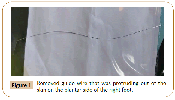
Eventually, the patient got cured after 6 days of hospitalization, the platelet count rose and she discharged. On the current admission to the surgery department of our institution, physicalexamination was unremarkable except for a guide wire that was present protruding out of the skin on the plantar side of the right foot (Figure 1).
An X-ray of right knee, X-ray PBH, X-ray right hip with proximal femur, X-ray right lower leg has been done.
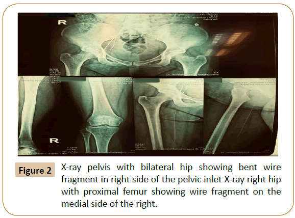
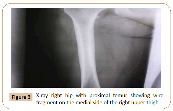
X-ray PBH, X-ray right hip with proximalfemur, showed abent radio opaque wire fragment on the right side of pelvic inlet measuring approximately 30-40 cm, presence ofanother wire fragment on themedial side of the right upper thigh, was also noted (Figures 2 and 3).
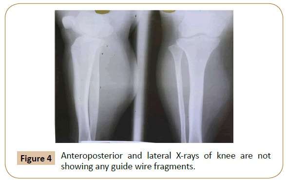
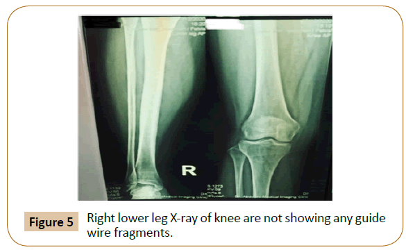
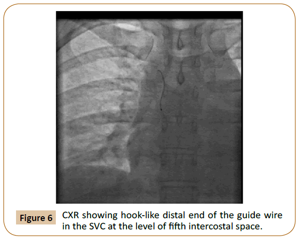
X-ray right knee, X-ray right lower leg did not report any guide wire fragment (Figures 4 and 5). Chest X-ray showed that the hook-like distal end of the guide wire (measuring approximately 4 cm) had broken off and extended upward travelling through the Inferior Vena Cava (IVC) and passed the right atrium and surprisingly finally terminated in the Superior Vena Cava (SVC) at the level of fifth intercostal space (Figure 6).
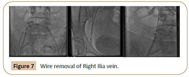
Surgical extraction of the guide wire fragments in the right Iliac vein and SVC, was performed in Cath lab. Under fluoroscopic guidance using a left femoral venous approach, the removal of the retained wire fragments was attempted with a Trefoil En- Snare by the interventional cardiology team.
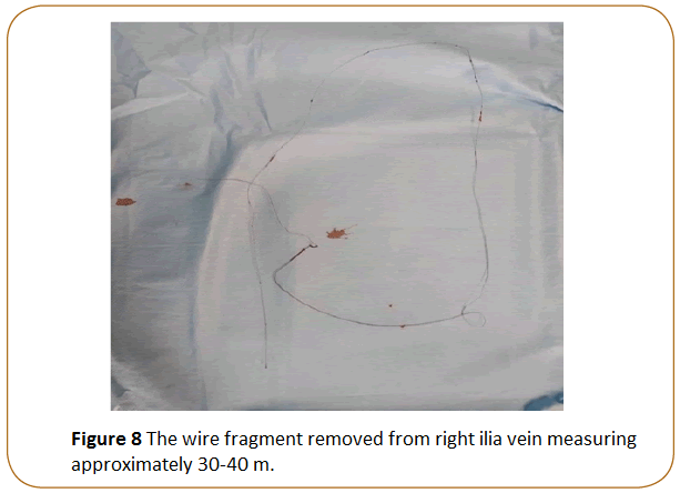
At first, the retained fragment in the right Iliac vein was removed. It was captured with the snare, which was guided from left femoralvein and was pulled out totally through the snare catheter, despite the weak adherence to vascular wall, measuring approximately 30-40 cm. (Surprisingly the length is more than a normal guide wire) (Figures 7 and 8).
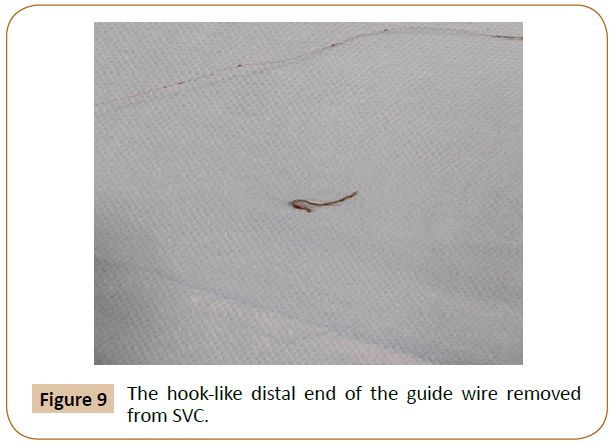
Secondly, the Trefoil En-Snare using the left femoral venous approach was guided toward the right atrium and the retained fragment in SVC was easily detached, measuring approximately 4 cm (Figure 9).
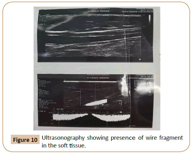
The fragment guide wire which has been in thigh, was not noticeable during venoangiography, according to that, we recognised it had been either in soft tissue orfemoralartery, and not in the femoralvein, So that ultrasonography wasdone. It showed that thefragment had been in the soft tissue (Figure 10).
Considering surgical therapy and discovering for the fragment would cause more damage, the patent was asked to wait.
The patent was then discharged home.
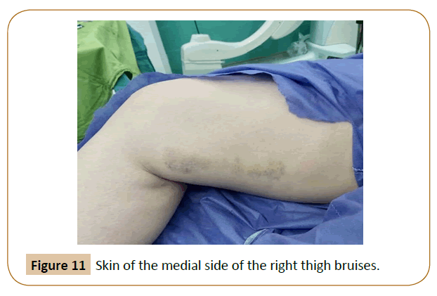
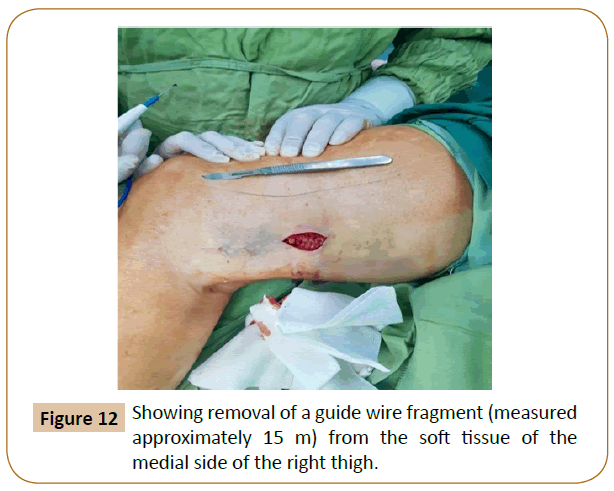
After one month, the fragment had been moved to the surface, skin surface was bruised and the wire could be touched under the skin (Figure 11). By the end, the last part (measured approximately 15 cm) was removed by a small incision (Figure 12).
Unbelievably, it was seen that the whole removed retained guide wire was about 60 cm, which is obviously more than the length of a normal guide wire that is about 20-25 cm, two theories were possible:
• Twocatheterwasused.
• Core wire and spring coil (components of guide wire) were detached and werestretched.
We noticed, the second theory was correct and the core wire was stuck in the right pelvic whereas the spring coil art was in soft tissue on the medial side of the right upper thigh.
Discussion
Guide wires are used in almost all intra vascular interventions, including central venous catheterisation.
There are many complications associated with guide wires insertion, a rare but dangerous complication of CVC placement is the guide wire retention which may result in vascular damage, thrombosis, embolism, infection, cardiac arrhythmias, cardiac tamponade, cardiac perforation [7,8]. Although the presented case had not developed any of these symptoms.
There are many riskfactorsthat can affectintra vascular guide wire retention, including experience of the operator, distraction during the procedure, high work load or lack ofproper supervision.
Lack of proper supervision is the commonest risk factor, with most of the cases seen with trainee doctors [9].
The incidence of guide wire retention, has been estimated at one per few thousands placements of CVC.
In New York Staten between 2008 and 2009, 80 cases of retained guide wire were reported, making them the most commonly reported non surgically retained stuff [10].
Most guide wires retentions are found immediately, usually during or shortly after the procedure. However, some may be found after a long period, as in ourcase (afterabout twoyears).
Detection of guide wire retention may also be incidental, postprocedure on radiographs whether performed immediately after the procedure or done later for other reasons [8].
In our patent guide wire detection occurred when it was emerged out through the skin to the outside of the body, which is an extremely rare occurrence. Complications related to wire retention when found out was pain.
Conditionof thewire fragments in the body(during the time of diagnosis or retrieval), a smallpiece of wire which was protruded through the skin about 2 years after CVC insertion.
a bent radio opaque wire fragment on the right side of pelvic inlet, which removed without breaking of wire. Another wire fragment in soft tissue on the medial side of the right upper thigh, which removed with a small incision through the skin.
The hook-like distal end of the guide wire which was embolised, and was terminated in the SVC, easily detached and removed without breaking of wire.
In this case, CVC insertion took part in the intensive care unit (ICU), which may have made it easier to miss the findingon CXR, due to the presence of multiple wires attached to patient’s body at that time.
In spite of following procedure protocols, guide wire retention does not decrease, as this depends on the human factor, as remembering to remove the guide wire [11]. To minimize the risk of guide wire retention, more stringent procedural protocol is needed, mariyaselvam et al. have investigated the use of a locked procedure kit, where the suture, suture holder and antimicrobial dressing are placed in a special pack where the guide wire acts as a key and is needed to open the pack [11].
This procedure requires that the clinician asserts that he has removed the guide wire before trying toplace the sutures,and hasbeenconfirmedtobe an effective preventative approach that improves patent safety [11].
In many cases, the guide wire removal is done through percutaneous endovascular means, using a goose neck snare device [12].
Conclusion
Guide wire retention is a rare though an increasingly reported complication due to increasing use of central line. To minimise such complications, performing these techniques with the guidance of skilled and experienced operators, is of great importance.
As most cases are found immediately during or following the procedure, rare cases are found late on X-rays. Presentations of retained guide wire by emerging out through the skin after years of insertion, is very unusual [13].
References
- Pokharel K, Biswas BK, Tripathi M, Subedi A (2015) Missed central venous guide wires: a systematic analysis of publishedcase reports. Crit Care Med 43: 1745-1756.
- Abuhasna S, Abdallah D, Rahman M(2011) The forgotten guide wire: a rare complicationof haemodialysis catheter insertion. J Clin Imaging Sci 5: 55-57.
- Billiar T, Andersen D, Hunter J, Brunicardi F, Dunn D, et al. (2004) Schwartz's principles of surgery. McGraw-Hill Professional 5: 314-342.
- Ruesch S, Walder B, Tramèr MR (2002) Complications of central venous catheters: internal jugular versus subclavian access-a systematic review. Crit care med30:454-560.
- Kornbaun C (2015) Central line complications. Int J CritIlln Inj Sci 5: 170-178.
- Guo H, Peng F, Ueda T (2006) Loss of the guide wire: a case report. Circulation journal: official. J Jpn Circ J 70:1520-1522.
- Amit A, Jyotsna S, Kasyap VK (2016) Retention of guide wire: A rare but avoidable complication of central venous catheterization. J Compr Pediatr7.
- Choudhary RS, Shah NJ, Audichya A, Gohil R (2020) A case of retained guide wire spontaneous emerging from skin near right knee 8 months after central venous catheterization. Asian J Case Rep Surg 28:10-15.
- Schummer W, Schummer C, Gaser E, Bartunek R (2002) Loss of the guide wire: mishap or blunder?.Br J Anaesth 88:144-146.
- PCI HR (2013)Anincidental complication of central line catheter placement: retention of a guide wire. Anaesth 21: 52-53.
- Mariyaselvam MZ, Catchpole KR, Menon DK, Gupta AK, Young PJ (2017) Preventing retained central venous catheter guide wires: A randomized controlled simulation study using a human factors approach. Anaesth 127: 658-665.
- Cheddie S, Singh B (2013) Guidewire embolism during central venous catheterization: Options in management. Int J Surg 30:1-4.
- Arnous N, Adhya S, Marof B (2019) A case of retained catheter guidewire discovered two years after central venous catheterization. Am J Case Rep 20:1427.
Open Access Journals
- Aquaculture & Veterinary Science
- Chemistry & Chemical Sciences
- Clinical Sciences
- Engineering
- General Science
- Genetics & Molecular Biology
- Health Care & Nursing
- Immunology & Microbiology
- Materials Science
- Mathematics & Physics
- Medical Sciences
- Neurology & Psychiatry
- Oncology & Cancer Science
- Pharmaceutical Sciences
